In Winter Quarter, I suffered my second concussion. While it was less severe than my third-grade concussion, I was still out of commission for over two months.
Despite the need for properly diagnosing dysfunction in the most important organ in the body – the brain – current concussion testing tools rely on patient effort or subjective reporting. For example, at Searle I would answer questions about my mood, whether I was experiencing headaches, sleeping habits, dizziness and other symptoms. Confounding factors, such as the fact that I was also suffering from an infection at the time of the concussion, could have affected the diagnostics, which highlights one of the key problems with subjective reporting. A more objective method of diagnostics is required to properly diagnose concussions, and such a method was presented at CommFest by Professor Nina Kraus, Director of the Auditory Neuroscience Laboratory, Tory Lindley, Northwestern’s Head Athletic Trainer and President-elect of the National Athletic Trainers’ Association, and Dr. Cynthia LaBella, professor of pediatrics at Feinberg School of Medicine and the medical director for the Institute for Sports Medicine at Lurie Children’s Hospital.
According to the speakers, there are over two million concussions in the US annually. At Northwestern, there are over 500 student-athletes covered by four primary care MDs, 24 athletic trainers and over 30 medical specialties and subspecialties. On a more local note, according to the speakers, 10 percent of NU football players suffer concussions per season. To ensure the safety of concussion sufferers, an independent neuro-consultant is employed to promote objective diagnoses. Nonetheless, because of the lack of objectivity in current testing diagnostics, the researchers asked the question: “What if we have a way to measure the brain objectively before the season and after the season?” This is achieved using frequency-following response (FFR), which uses three electrodes to detect biological markers of concussions in real-time through how electrical waves in the brain are measured as patients listen to and comprehend sound. According to Professor Kraus, “Electricity is the currency of the nervous system,” which means that each individual has their own neural signature that is detected by FFR.
In the studies conducted using sound processing to evaluate concussions, FFR identified 90 percent of concussed individuals and cleared 95 percent of the control group. For the 40 NU football student-athletes affected by concussions in the 2016 and 2017 seasons, timing, pitch and timber had widespread disruption from preseason FFR data. This is potentially due to fatigue in the brain.
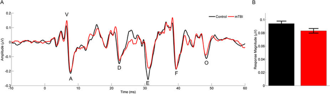
Brain Responses are Smaller in the Concussed Children (Red Line)
Photo Courtesy of Nature
In addition to being an objective diagnostic of concussions, the FFR tool is repeatable. This allows for it be run multiple times on the same individual, and the non-invasive nature of the tool is a huge plus. MRIs and CT scans are sometimes employed by medical personnel to rule out other conditions such as brain hemorrhaging or skull fractures, but concussions do not appear in such scans, but the radiation that hits patients’ bodies certainly does remain.
“It’s not so much the nature of the brain injury, but the nature of the brain that is injured,” said Dr. LaBella. This means that how the injury occurs that matters, but the results of such an injury on the brain. With this new diagnostic tool, the athletes and the general population can look forward to more comprehensive concussion diagnostics and better ways to ensure that concussion-free versus concussed is not a matter of whether you are a two or a three out of ten on a dizziness scale.